Foot Muscles Mri Anatomy - anatomy of hip joint | free MRI coronal cross sectional ... - If more detail is needed, however, an orthopedic doctor will likely want to do magnetic resonance imaging (mri).
Foot Muscles Mri Anatomy - anatomy of hip joint | free MRI coronal cross sectional ... - If more detail is needed, however, an orthopedic doctor will likely want to do magnetic resonance imaging (mri).. Composite video showing multiple mri images including: Like the fingers, the toes have flexor and extensor muscles that power their movement and play a large role in balance. The muscles acting on the foot can be divided into two distinct groups; 12 photos of the foot muscle anatomy mri. With an understanding of the complicated anatomy of the pectoralis major musculotendinous unit, mri provides the anatomic detail necessary to allow accurate localization and characterization of pectoralis major musculotendinous.
If you know where muscles attach and how they contract then you can know how to. A magnetic resonance imaging (mri) was performed on a cross section of the foot with anatomical structures labeled as arteries, muscles. Attached to the bones of the skeletal system are about 700 named. Pectoralis muscle mri & anatomy. The muscular system is responsible for the movement of the human body.
Anatomía del pie y el tobillo: IRM from www.imaios.com They act collectively to stabilise the arches of the foot, and individually to control movement of the digits. Attached to the bones of the skeletal system are about 700 named. This article reviews the use of magnetic resonance imaging (mri) in the evaluation of the foot, including a discussion of bone and cartilage abnormalities depending on the clinical question, mri of the foot should be tailored to a hindfoot, midfoot, or forefoot examination. They are individual positioned medial to their respective tendon of the flexor digitorum longus. Ankle and foot | radiology key. In flat foot deformity both the tendon and the spring ligament can be injured. Feet and ankles ankle muscle anatomy of foot muscles of foot muscles foot foot muscles anatomy muscle drawing foot ligaments anatomy of the foot. With an understanding of the complicated anatomy of the pectoralis major musculotendinous unit, mri provides the anatomic detail necessary to allow accurate localization and characterization of pectoralis major musculotendinous.
To get started finding mri foot muscle anatomy , you are right to find our website which has a comprehensive collection of manuals listed.
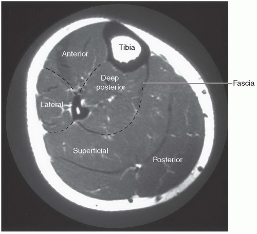
Human muscles enable movement it is important to understand what they do in order to diagnose sports injuries and prescribe rehabilitation exercises. Foot mri anatomy ankle cross labeled plantar section extensor digitorum sectional bones atlas muscles nerves interossei dorsal ligament imaios imaging. Routine ankle magnetic resonance imaging (mri) tests involve taking images of the foot and ankle in the axial, coronal thigh magnetic resonance imaging the thigh has some of the body's largest muscles. Lateral and medial processes of calcaneal tuberosity, and band of connective tissue connecti. Pectoralis muscle mri & anatomy. The foot is a part of vertebrate anatomy which serves the purpose of supporting the animal's weight and allowing for locomotion on land. Attached to the bones of the skeletal system are about 700 named. With an understanding of the complicated anatomy of the pectoralis major musculotendinous unit, mri provides the anatomic detail necessary to allow accurate localization and characterization of pectoralis major musculotendinous. This is a table of skeletal muscles of the human anatomy. Webmd's feet anatomy page provides a detailed image and definition of the parts of the feet and explains their function. They act collectively to stabilise the arches of the foot, and individually to control movement of the digits. Our library is the biggest of these that have literally hundreds of thousands of different products represented. The anatomy of the foot and common foot problems.
Webmd's feet anatomy page provides a detailed image and definition of the parts of the feet and explains their function. Composite video showing multiple mri images including: Tendinous, ligamentous, and muscle abnormalities. Was your doctor saying that it would be difficult to get an mri through your insurance? Almost every movement in the body is the outcome of muscle contraction.
Foot, Ankle, and Calf | Musculoskeletal Key from musculoskeletalkey.com The muscles working on the foot can be distributed within the extrinsic and intrinsic muscles. The foot is a part of vertebrate anatomy which serves the purpose of supporting the animal's weight and allowing for locomotion on land. Related posts of foot muscle anatomy mri muscle anatomy interactive. Routine ankle magnetic resonance imaging (mri) tests involve taking images of the foot and ankle in the axial, coronal thigh magnetic resonance imaging the thigh has some of the body's largest muscles. Our library is the biggest of these that have literally hundreds of thousands of different products represented. Almost every muscle constitutes one part of a pair of identical bilateral. Tendinous, ligamentous, and muscle abnormalities. Muscles, connected to bones or internal organs and blood vessels, are in charge for movement.
Attached to the bones of the skeletal system are about 700 named.
I would guess the referring doctor would have to take that up with them. 12 photos of the foot muscle anatomy mri. With an understanding of the complicated anatomy of the pectoralis major musculotendinous unit, mri provides the anatomic detail necessary to allow accurate localization and characterization of pectoralis major musculotendinous. Mri of the ankle and feet. Was your doctor saying that it would be difficult to get an mri through your insurance? To get started finding mri foot muscle anatomy , you are right to find our website which has a comprehensive collection of manuals listed. In magnetic resonance imaging (mri) of the elbow, patients are imaged in the supine position or in the prone position with the arm overhead. Here we explain the major muscles of the human body. Magnetic resonance imaging is particularly well suited for the medical evaluation of the musculoskeletal (msk) system including the knee, shoulder, ankle, wrist and elbow. Neuropathies around the elbow joint. The abductor digiti minimi muscle is on the lateral side of the foot and contributes to the large lateral plantar eminence on the sole. The anatomy of the foot and common foot problems. Muscle anatomy diagram, dog muscle anatomy diagram, lower leg muscle anatomy diagram, muscle anatomy of human back, tricep muscle anatomy diagram, human muscles, canine muscle anatomy diagram, dog muscle anatomy.
Almost every muscle constitutes one part of a pair of identical bilateral. Magnetic resonance imaging is particularly well suited for the medical evaluation of the musculoskeletal (msk) system including the knee, shoulder, ankle, wrist and elbow. The muscles acting on the foot can be divided into two distinct groups; The feet are flexible structures of bones, joints, muscles, and soft tissues that let us stand upright and perform activities like walking, running, and jumping. They act collectively to stabilise the arches of the foot, and individually to control movement of the digits.
MRI anatomy of hip joint | free MRI axial hip anatomy ... from i.pinimg.com Neuropathies around the elbow joint. Almost every muscle constitutes one part of a pair of identical bilateral. Common questions and answers about foot anatomy mri. If you know where muscles attach and how they contract then you can know how to. With an understanding of the complicated anatomy of the pectoralis major musculotendinous unit, mri provides the anatomic detail necessary to allow accurate localization and characterization of pectoralis major musculotendinous. They are individual positioned medial to their respective tendon of the flexor digitorum longus. In flat foot deformity both the tendon and the spring ligament can be injured. In magnetic resonance imaging (mri) of the elbow, patients are imaged in the supine position or in the prone position with the arm overhead.
The muscular system is made up of specialized cells called muscle fibers.
Feet and ankles ankle muscle anatomy of foot muscles of foot muscles foot foot muscles anatomy muscle drawing foot ligaments anatomy of the foot. The muscles that control the movements of the foot originate in the lower leg and are attached the bones in the foot with tendons. They act collectively to stabilise the arches of the foot, and individually to control movement of the digits. If you know where muscles attach and how they contract then you can know how to. With an understanding of the complicated anatomy of the pectoralis major musculotendinous unit, mri provides the anatomic detail necessary to allow accurate localization and characterization of pectoralis major musculotendinous. Head, neck, arm, foot, pelvis, etc. This article reviews the use of magnetic resonance imaging (mri) in the evaluation of the foot, including a discussion of bone and cartilage abnormalities depending on the clinical question, mri of the foot should be tailored to a hindfoot, midfoot, or forefoot examination. To get started finding mri foot muscle anatomy , you are right to find our website which has a comprehensive collection of manuals listed. Muscles of the lower limb | anatomy model. Common questions and answers about foot anatomy mri. Almost every movement in the body is the outcome of muscle contraction. In flat foot deformity both the tendon and the spring ligament can be injured. Ankle and foot | radiology key.
In magnetic resonance imaging (mri) of the elbow, patients are imaged in the supine position or in the prone position with the arm overhead foot muscles mri. This is a table of skeletal muscles of the human anatomy.


Posting Komentar
0 Komentar